Hemoglobin | Complete Guide
Last updated on December 14th, 2023
What is Hemoglobin?
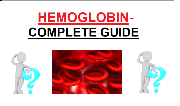
Hemoglobin (spelled hemoglobin and abbreviated HB or HGB) is a metalloprotein transporting iron-rich oxygen found in red blood cells of vertebrates and in the tissues of some insatiable. The hemoglobin in the blood transports oxygen from the lungs or gills to the rest of the body (ie tissue), where it releases oxygen for use by cells.
In mammals, protein is composed of about 97% of the dry red blood cells and about 35% of the total (including water).
The oxygen binding capacity of hemoglobin is between 1.36 and 1.37 ml O2 per gram of hemoglobin, which increases the total blood oxygen capacity by seventy times.
Hemoglobin is also found outside the red blood cells and progenitor lines that produce them. Other hemoglobin-containing cells include the A9 dopaminergic neuron, macrophages, alveolar cells, and mesangial cells of the kidneys of the substantial nigra. The role of hemoglobin in these tissues is an antioxidant and a regulator of iron metabolism rather than oxygen transport.
Hemoglobin | History of research:
The oxygen-carrying protein hemoglobin was discovered by Henfield in 1840. In 1851 Otto Funk published a series of articles in which he slowed down the hemoglobin crystals by evaporating the solution with a protein solution after diluting the red blood cells with the help of solutions such as pure water, alcohol, or ether. Told about growing.
The role of hemoglobin in blood was given by physiologist Claude Bernard. The name hemoglobin is derived from the combination of the terms heme and globin, suggesting that each subunit of hemoglobin is a globular protein in the heme group. Each heme group has one iron atom, which can bind an oxygen molecule through ion-induced forces. The most common hemoglobin of mammals has four such subunits.
Genetics:
Hemoglobin consists mostly of proteins (globin chains) and these proteins are made up of chains of amino acids. These chains are linear in form, such as a written sentence containing letters or beads in a garland. The variation in the types of amino acids in the protein chain of amines acids in all proteins determines the chemical properties and function of that protein. The same applies to hemoglobin, in which a series of amino acids can affect important functions such as the attraction of proteins to oxygen.
The amino acid chains of globin proteins in hemoglobin differ among different species, although the differences increase with the distance of growth between species. For example, the most common hemoglobin chains in humans and chimpanzees are similar, while this same chain differs from the most common amino acid chain of guerrillas by only one amino acid in the alpha and beta-globin protein chains. These differences are more in castes with less affinity. Like proteins other than hemoglobin, differences in DNA chains between species are greater than differences in amino acid chains coded by them, as different DNA chains may point to the same amino acid.
In any one species, different types of hemoglobin are always present, although one chain is usually the most common in every species. Variations of hemoglobin protein genes generate different types of hemoglobin. There is no disease due to many of these deformed types. But some malformed hemoglobins cause a group of genetic diseases called hemoglobinopathies. Sickle cell disease is the most famous hemoglobinopathy, which is the first human disease, the process of which is understood at the molecular level. In one (mostly) group of other diseases, thalassemia, small amounts of normal or sometimes abnormal hemoglobins are produced due to problems and pathologies of the globin gene control. All these diseases cause anemia.
Like other proteins, chains of hemoglobin’s amino acids can be adaptive. For example, recent studies have shown how deer mice living in the mountains survive the thin air found at heights with the help of their different gene types. A researcher at the University of Nebraska-Lincoln found malformations in four different genes that are thought to be responsible for the differences between deer mice living in low-lying areas and mountains. Examining wild bees captured from the mountains and lowlands, it was found that the genes of both breeds were identical, except for those that control their hemoglobin’s oxygen-carrying capacity. The genetic variation of mountain rodents gives them the ability to make better use of oxygen, as they are available in small quantities at high places such as mountains. Hemoglobin deformities of ancient elephants also enabled oxygen supply in low-temperature areas, allowing them to live at higher elevations during the Pleistocene.
Synthesis:
Synthesis of hemoglobin occurs in a complex step chain. The synthesis of the heme portion occurs in various steps in the mitochondria and cytosol of immature red blood cells, while the globin protein portion is synthesized by ribosomes in the cytosol. Hemoglobin in the bone marrow is produced in the cell throughout the period of its early development from the proerythroblast to the reticulocyte stage. At this time the nucleus of mammals’ red blood cells disappears, but this does not happen in birds and many other species. Despite the absence of a nucleus in mammals, the remaining ribosomal RNA produces hemoglobin until the reticulocyte’s RNA is depleted after entering the bloodstream. It has got its fake form and name).
Formation:
Hemoglobin exhibits properties of both tertiary and quartile compositions of proteins. Most of the amino acids of hemoglobin form alpha helices that are associated with small phallic segments. Hydrogen bonds stabilize helical sections within this protein, causing attraction within the molecule and each polypeptide chain is twisted into a characteristic shape. The quaternary structure of hemoglobin is due to its four subunits located in the tetrahedral system.
In most humans, a hemoglobin molecule is a group of four globular protein subunits. Each subunit is made up of a protein chain tightly connected to a nonprotein heme group. Each protein chain is arranged in a globin fold arrangement as a set of fused alpha-helix conformational segments, as other heme/globin proteins such as myoglobin have the same folding motif-like arrangement. This folding pattern has a place, which holds the heme group firmly.
The heme group contains an iron ion that resides in a heterocyclic ring, called porphyrin. This porphyrin ring consists of four pyrrole molecules, which are attached to the iron ion at the center as a circle. The iron ion, which is the location of the oxygen bond, works together with four nitrogens living in the same plane at the center of the ring. The iron is strongly attached to the globular protein via the imidazole ring of the F8 histidine residue located below the porphyrin ring. A hinge can reversibly bind oxygen via a coordinate covalent bond, thus completing the octahedral group of six ligands. Oxygen binds in an end-on bent geometry, where the oxygen atom is bound to iron and the other exits at an angle. When oxygen is not bound, a water molecule with a weak bond fills that space, creating a distorted octahedron.
Although carbon dioxide is transported by hemoglobin, it does not compete with oxygen for iron-binding sites but is actually linked to the protein chains of the structure.
Iron ions can occur in the Fe2 + or Fe3 + state, but ferric hemoglobin (methemoglobin) (Fe3 +) cannot bind oxygen. Oxygen in binding temporarily and reversibly oxidizes (Fe2 +) to (Fe3 +), while oxygen is temporarily converted to superoxide. In this way, iron has to be in the oxidation state to bind oxygen. If the superoxide anion corresponding to Fe3 + is protonated, the hemoglobin iron remains oxidized and unable to bind oxygen. In such a situation the enzyme methemoglobin reductase may eventually reactivate methemoglobin by decreasing the iron center.
The most common hemoglobin type in adult humans is a tetramer (containing four subunit proteins) called hemoglobin A, which consists of two alpha and two beta subunits with non-covalent bonds composed of 141 and 146 amino acids, respectively. This is indicated by α2β2. These subunits are anatomically similar and approximately the same size. The molecular weight of each subunit is about 17000 daltons and the total molecular weight of the tetramer is about 68000 daltons (64,458 g / mol). In this way 1 g / dL = 0.1551 millimole / l. Hemoglobin A is the most refined molecule among hemoglobin molecules.
In human babies, the hemoglobin molecule is made up of 2 alpha and 2 gamma chains. As the baby grows, the gamma chains are gradually replaced by beta chains.
These four polypeptide chains are bound together by salt bridges, hydrogen bonds, and hydrophobic interactions. There are two types of interactions between alpha and beta chains – α1β1 and α1β2.
Broadly, hemoglobin can be saturated (oxyhemoglobin) or unsaturated (deoxyhemoglobin) from oxygen molecules. Oxyhemoglobin originates during physiological respiration when the heme portion of the protein hemoglobin in red blood cells binds to oxygen. This process occurs in the pulmonary capillaries located near the alveoli in the lungs. Oxygen then reaches the cells through the bloodstream where it is used for the hydrolysis of aerobic glycolysis and oxidative phosphorylation in the formation of ATP. But it is not helpful in preventing blood pH deficiency. Respiration can reverse this condition by removing carbon dioxide, which causes the pH to rise back.
Deoxyhemoglobin is a type of hemoglobin devoid of bound oxygen. The absorptive spectrum of oxyhemoglobin and deoxyhemoglobin is different. Oxyhemoglobin has a much lower absorption of 660 nm wavelength than deoxyhemoglobin, while its absorption is slightly higher at 940 nm. For this reason, the color of hemoglobin is red and the color of deoxyhemoglobin is blue. This variation is used to measure the amount of oxygen in the patient’s blood by a device called a pulse oximeter.
Oxidation states of iron in oxyhemoglobin:
The oxidation state of oxidized hemoglobin is difficult to determine because by experimental measurements oxyhemoglobin (Hb-OTu) is diamagnetic (there are no unpaired electrons), yet low-energy electron structures in both oxygen and iron are paramagnetic (ie this compound Has at least one unpaired electron). The following are the minimum-energy types of oxygen and related iron oxidation conditions:
- Triple-oxygen, a species of low-energy oxygen, has two unpaired electrons in anti-binding π * molecular orbital.
- Iron (II) occurs in a high-spin structure where unpaired electrons are in the anti-Eg binding arbitral.
- Iron (III) has a paired number of electrons and therefore it is necessary to have one or more unpaired electrons in any energy state.
All these structures are paramagnetic (with unpaired electrons), rather than diamagnetic. Thus, in order to understand the observed diamagnetism and unpaired electrons, an uneven distribution of electrons in the combination of iron and oxygen is necessary.
There are three possibilities for the production of diamagnetic (without net spin) HB-OTO:
- Low-spin Fe2 + is bound to singlet oxygen. Both low-spin iron and singlet oxygen are diamagnetic. However, the singlet type of oxygen is the higher-energy type of molecule.
- Bonding of low-spin Fe3 +. O2- (superoxide anion) and two unpaired electrons pair in a ferromagnetic manner that has diagenetic properties.
- Low spin Fe4 + is bonded to peroxide, O22-. Both are diamagnetic.
Straightforward information:
According to X-ray photoelectron spectroscopy, the oxidation state of iron is about 3.2.
According to the infrared frequencies of the O – O bonds, the length of the dam is compatible with the superoxide (bond order about 1.6, and superoxide 1.5).
In this way, the closest formal state of iron in Hb-OTu is the oxidation +3 state, with the oxygen-1 state (as superoxide. O2-). The diamagnetism in this structure arises from the single unpaired electrons on the superoxide arranged in iron in a counter-magnetogenetically manner from the single unpaired electrons on the iron, causing the entire structure to receive no total spin, according to the diamagnetic oxyhemoglobin of the experiment.
It is not surprising to find that the second of the three possibilities given above for diamagnetic oxyhemoglobin is correct by experiment – both singlet oxygen (possibility No. 1) and large division of charge (probability No. 3) are unfavorable high-energy conditions. Moving to a higher oxidation state of iron in HB-OTO reduces the size of AMU, allowing it to move to the bottom of the peripherin ring, and initiating the allosteric changes observed in globulins by pulling the coordinated histidine residue.
According to preliminary theories presented by bio-inorganic chemists, probability no. 1 was correct and the oxidation condition to iron II. Should be in This was thought to be because iron’s oxidation state in the form of methemoglobin when superoxide is not present to “hold” the .O2-electron, makes hemoglobin inefficient to bind the normal triplet O2 in the air. Therefore, it is assumed that when oxygen gas is bound in the lungs, iron remains in the form of Fe (II). The chemistry of iron in this earlier classic model was intriguing, but the need for diagenetic high-energy singlet oxygen was never understood. It was argued that the binding of the oxygen molecule to high spin iron (II) was octahedral of strong-field ligands. Keeps it in the field – due to this change in the field, the increase in the crystal area causes the energy to be split, due to which the electrons of iron The loops form a low-spin structure, which is diamagnetic in Fe (II). This forced low-spin pairing occurs on oxygen binding but is not sufficient to explain the change in iron size. In order to understand the small size and increased oxidation state of iron, and the weak oxygen bond, it is necessary to remove an extra electron from the iron by oxygen.
It should be kept in mind that the determination of the whole-number oxidation state is a formalism since covalent bonds are not necessary for flawless bond sequences in full electron transfer. In this way, all three models of paramagnetic HB-OTUs may be responsible to some extent for the actual electronic structure of HB-OTUs. But it seems more correct to have Fe (III) of the iron model in Hb-OTu than Fe (II).
Types in humans:
Different types of hemoglobin are a part of the normal development of the fetus and baby, but there may also be morbid deformable types of hemoglobin produced due to differences in genetics in a population. Some famous hemoglobin types such as sickle-cell anemia are responsible for diseases and are called hemoglobinopathy. Other types do not cause any special sickness, so they are called degenerative types.
In the fetus –
- Gower 1 (ζ2ε2)
- Gower 2 (α2ε2) (PDB 1A9W)
- Hemoglobin portland (ζ2γ2)
In baby:
- Hemoglobin F (α2γ2) (PDB 1FDH)
In adults –
- The Hemoglobin A (α2β2) – the most common, greater than 95%
- Hemoglobin Etu (α2δ2) – δ chain synthesis begins in the third trimester and its normal levels in adults are 1.5–3.5%.
- Hemoglobin F (α2γ2) – In adults, hemoglobin F occurs only in a limited number of red cells called F-cells. But HB F levels may increase in people with sickle-cell anemia.
Disease-causing types –
- The Hemoglobin H (β4) – a type of hemoglobin generated by a tetramer of β chains, which can be found in types of α thalassemia.
- Hemoglobin Barts (γ4) – a type of hemoglobin generated by a tetramer of γ chains that can be found in variants of α thalassemia.
- Hemoglobin S (α2βS2) – a type of hemoglobin found in sickle cell patients. It differs in the β-chain gene, which alters the properties of hemoglobin and cycling of red blood cells.
- Hemoglobin C (α2βC2) – another type generated due to changes in the β-chain. This type causes a mild long-term hemolytic anemia.
- Hemoglobin E (α2βE2) – another type generated due to changes in the β-chain. This type causes a mild long-term hemolytic anemia.
- Hemoglobin is a type of AS-sickle cell tract that contains an adult gene and a sickle cell gene.
- Hemoglobin SC disease – another type with a sickle cell gene and another hemoglobin C gene.
Vertebrate animals weathering:
When red cells reach the end of their lives due to age or disorders, they burst, the hemoglobin molecule splits and iron is reused. When the porphyrin ring disintegrates, its residues are normally secreted into the bile by the liver. This process also produces one carbon monoxide molecule for every molecule of heme. It is one of the few sources of carbon monoxide production in the human body and is responsible for the normal blood levels of carbon monoxide in people breathing in pure air. The second main product of heme weathering is bilirubin. If red blood cells are getting destroyed faster than normal, increased levels of this chemical can be found in the blood. Incorrectly weathering hemoglobin protein or hemoglobin released from blood cells can rapidly block small blood vessels, especially the delicate blood vessels that drain the kidneys, causing damage to the kidneys.
Role in diseases:
Deficiency of hemoglobin may result in a decrease in the number of hemoglobin molecules, such as in anemia, or a decrease in the ability of each molecule to bind oxygen at a partial pressure equal to oxygen. Hemoglobinopathies (the abnormal composition of hemoglobin resulting from genetic disorders) can be the cause of both. In any case, the lack of hemoglobin decreases the ability of the blood to carry oxygen. In general, hemoglobin deficiency should be distinguished from a lack of oxygen in the blood, or a decrease in the partial pressure of oxygen in the blood, although both are involved in the causes of hypoxia (insufficient oxygen gain to tissues).
Other common causes of low hemoglobin include blood loss, nutritional deficiency, blood marrow problems, chemotherapy, renal failure, or abnormal hemoglobin (such as in sickle cell disease).
High places, smoking, dehydration, or tumors can cause high hemoglobin levels.
The oxygen-carrying capacity of each hemoglobin molecule is normally modified by altered blood pH or carbon dioxide, creating a transformed oxygen – hemoglobin dissolution curve. It can also be replaced by diseases, eg carbon monoxide poisoning.
Symptoms of anemia are caused by a decrease in the amount of hemoglobin, with or without a deficiency of red blood cells. There are many reasons for anemia, although an iron deficiency in the western world and iron deficiency anemia arising from it are the main reasons. Because heme synthesis is reduced due to the absence of iron, red blood cells appear to be light red in color (lack of hypochromic-red hemoglobin pigment) and small in size (microcytic) in iron-deficiency anemia. Another anemia is rare. In hemolysis (more rapid loss of red blood cells) the hemoglobin metabolites bilirubin cause jaundice, and the hemoglobin flowing into the blood can cause the kidneys to fail.
Some disorders of the globin chain are associated with hemoglobinopathies, such as sickle cell disease and thalassemia. Other disorders, as stated at the beginning of this article, are mild and are described only as hemoglobin types.
Errors of metabolic pathways of heme synthesis are found in a group of genetic diseases called porphyria. King George III of the United Kingdom was possibly the most famous patient of the disease.
To a lesser extent, hemoglobin A is slowly combined with glucose at the valene terminal (an alpha-amino acid) of each beta chain. The molecule obtained from this is often called HbA1c. As the amount of glucose in the blood increases, the percentage of HbA changing to HbA1c also increases. Patients with diabetes who have high glucose levels also have higher HbA1c. The percentage of HbA1c represents the average glucose levels in the blood over a longer period of time (half-life of red blood cells, which is 50–55 days), as the rate of HBA combined with glucose is slower.
Glycosylated hemoglobin is a type of hemoglobin that is combined with glucose. Glucose is combined with hemoglobin throughout the life of red blood cells, which is about 120 days. Glycosylated hemoglobin levels are measured to monitor long-term control of type 2 diabetes (Ta2DM) disease. Glycosylated hemoglobin levels in red blood cells are increased if Ta2DM is not controlled properly. Generally, it should be around 4-5.9%. 7% for patients with T2DM, even when it is difficult. It is advisable to keep less than .9%. Levels greater than 12% signify poor control of glycosylated hemoglobin, and 12%. Exceeding levels are considered very bad control. Patients whose glycosylated hemoglobin level is 7%. The chances of having complications due to diabetes are significantly reduced (compared to those in which it is 8%. Or higher).
The research was carried out to examine the effect of two different types of exercise programs (combined aerobic and anti-exercise programs and aerobic exercise only) on glycosylated hemoglobin levels of patients with T2DM –
In this investigation, total glucose control was measured as glycosylated hemoglobin (HbA or A). Glucose control was better than aerobic only when resistance exercise was combined with aerobic exercise. 0.8% of glycosylated hemoglobin in the average effect of exercise programs. Included shortages that were similar to long-term diet and drug or insulin treatment results (0.6–0.8%. Reduction).
Increased hemoglobin levels are associated with a greater number or size of red blood cells, known as polycythemia. This increase is due to congenital heart disease, cor pulmonary, pulmonary fibrosis, too much erythropoietin, or polycythemia vera.
A study of a yoga practice called Yoga Nidra for half an hour every day showed an increase in hemoglobin levels.
Clinical use:
Hemoglobin content is one of the most commonly used blood tests and is commonly used as part of an overall blood count. E.g. It is tested before or after blood donation. The results are expressed in grams per liter, grams per dL, or moles per li. 1 gram per dL equals about 0.15 millimole per liter. Common levels are:
- Male: 13.8 to 18.2 g / dL (138 onto 182 g / L, or 2.15 mM to 2.8 mM (mmol / L))
- Women: 12.1 to 15.1 g / dL (121 to 151 g / L, or 1.89 mM to 2.35 mM)
- Children: 11 to 16 g / dL (111 to 160 g / L, or 1.73 mM to 2.5 mM)
- Pregnant women: 11 to 12 g / dL (110 to 120 g / L, or 1.73 mM to 1.89 mM)
Normal levels of hemoglobin should be at least 11 g / dL in the first and third trimesters of pregnant women and at least 10.5 g / dL in the second trimester.
If the quantity is less than normal, it is called anemia. Anemia is categorized according to the size of red blood cells(RBC). If red cells are small, anemia is microcytic, if large, macrocytic, otherwise called normocytic.
The hematocrit, the ratio of red blood cells to the volume of blood, is usually about three times the hemoglobin level. E.g. If the amount of hemoglobin is 17, the hematocrit will be 51.
Long-term control of blood glucose levels can be done by measuring the amount of HbA1c. Samples are needed to directly measure this, as blood sugar levels can vary greatly throughout the day. HbA1c is the result of the reaction of hemoglobin A to glucose. HbA1c is high when glucose is high. As this reaction is slow, the ratio of HbA1c represents the average glucose levels in the blood over the half-life (50–55 days) of red blood cells. A ratio of 6.0 percent or less of HbA1c is considered to be a good long-term control of glucose, while greater than 7.0 percent. The level is considered insufficient control. This test is especially useful for patients with diabetes.
STAY HAPPY, STAY HEALTHY!!
Also Read:
- Most Common Questions on Hemoglobin Levels

- Myths about increasing Hemoglobin Levels

- Treatment for High Hemoglobin Levels: How to Lower Your Levels Safely

To know more about Hemoglobin Levels, visit Hemolevel.com
